
A successful lung transplant in the operating room is only the start. The bronchial anastomosis - the surgical join where the donor airway meets the recipient’s - depends initially on collateral blood supply from the pulmonary artery, with full bronchial circulation taking weeks to re-establish. During that vulnerable window, anything from infection to immunosuppression to technical issues can produce one of a small family of airway complications. Recognising them early, and intervening with the right tool, often makes the difference between a stable graft and a failing one.
Why this matters
Airway complications affect roughly 10-15 % of lung transplant recipients. At their worst they look like rejection and are missed for weeks. At their best they are caught at a routine surveillance bronchoscopy and treated in a single 30-minute outpatient procedure.
The complications we look for
In transplant practice, five airway problems account for the vast majority of post-operative interventional pulmonology work:
1. Anastomotic dehiscence
A partial breakdown of the surgical join, usually appearing in the first 4 weeks. Presents with pneumomediastinum, persistent air leak, or pleural air-fluid level. Minor dehiscence may heal on its own with conservative management; larger defects may need covered stenting to bridge the gap while the tissue heals.
2. Anastomotic stenosis
The most common late complication, typically appearing 2-6 months after surgery. Granulation tissue, fibrosis, or ischaemic remodelling progressively narrow the airway. The patient may notice a drop in FEV1, new breathlessness on exertion, or wheeze that doesn’t respond to bronchodilators. Most cases respond well to a stepwise approach: balloon dilatation first, with cryotherapy or electrocautery for exuberant granulation, and stenting reserved for recurrent or resistant cases.
3. Bronchomalacia
Dynamic collapse of the airway, especially during forced expiration or cough. Often mistaken for asthma. Diagnosed at dynamic bronchoscopy with the patient awake at the end of the procedure or on expiratory CT. Treatment is more nuanced - some cases respond to non-invasive ventilation, others need a silicone or hybrid stent to maintain patency.
4. Endobronchial infection and colonisation
The transplanted lung is denervated and has no cough reflex below the anastomosis, which predisposes to bacterial colonisation (especially Pseudomonas, Staphylococcus aureus) and fungal overgrowth (Aspergillus, Candida). Surveillance bronchoscopy with bronchoalveolar lavage and biopsy is the only reliable way to detect and treat these early, often before symptoms appear.
5. Granulation tissue and exophytic lesions
Exuberant healing produces polyps and granulation tissue that can obstruct the lumen. Cryotherapy is the procedure of choice - it ablates the lesion with minimal collateral damage and produces cleaner re-epithelialisation than cautery in transplanted tissue.
The intervention toolkit
Modern interventional pulmonology gives us a graded set of techniques. The principle is simple: use the least invasive option that achieves a durable result.
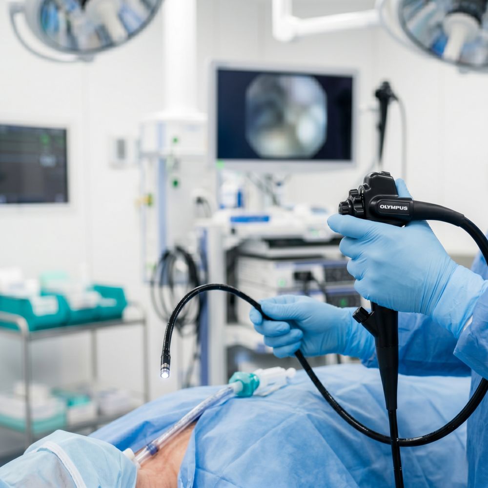
- Flexible bronchoscopy - the foundation. Used for diagnosis, biopsy, suction, lavage, and surveillance. Performed monthly or quarterly in the first year, more often if needed.
- Rigid bronchoscopy - the workhorse for therapeutic interventions. Provides a stable airway under general anaesthesia, allows simultaneous ventilation, and accommodates larger instruments. Essential for stenting, debulking, and managing major bleeding.
- Balloon dilatation - first line for symptomatic stenosis. A controlled-radial-force balloon is inflated across the narrowing for 60-90 seconds. Often needs 2-3 sessions but avoids the long-term issues of stents.
- Cryotherapy - freezes tissue rapidly to ablate granulation, polyps, mucus plugs, or blood clots. Preferred over cautery in transplant airways because it preserves the cartilaginous framework.
- Electrocautery and laser - reserved for cases where bleeding control matters or cryotherapy is insufficient. Used cautiously in transplanted tissue.
- Airway stenting - silicone (removable), self-expanding metallic (permanent or semi-permanent), or hybrid (uncovered metallic). The choice depends on lesion type, location, expected duration, and the patient’s overall trajectory.
Stents are powerful tools but each one introduces its own risks. The goal is always to leave the airway as close to its native geometry as possible.
A typical case - how a stenosis is managed
A patient 3 months post-bilateral lung transplant reports a 200 mL drop in FEV1 over 4 weeks and new dyspnoea climbing stairs. CT shows nothing dramatic. Bronchoscopy reveals 60 % narrowing of the right main bronchial anastomosis with surrounding granulation tissue.
The plan unfolds in three steps:
- First session: rigid bronchoscopy under general anaesthesia. Cryotherapy applied to the granulation tissue, followed by balloon dilatation to 12 mm. The patient goes home the same day. FEV1 recovers 150 mL over the next week.
- Re-evaluation at 4 weeks: partial recurrence. Second session of dilatation, lumen now stable at 10 mm.
- Re-evaluation at 3 months: stable, no recurrence. Surveillance continues every 6 months without further intervention.
Most stenoses are managed this way - without ever needing a stent. Stents are powerful tools but each one introduces its own risks: migration, granulation, mucus impaction, and the small but real risk of pressure necrosis. The goal is always to leave the airway as close to its native geometry as possible.
When the airway needs a stent
Stenting becomes necessary when:
- The stenosis recurs within 4-6 weeks despite repeated dilatation
- The airway shows dynamic collapse (malacia) that does not respond to non-invasive ventilation
- There is dehiscence requiring a covered stent to bridge the defect
- There is extrinsic compression from a haematoma, abscess, or post-surgical fibrosis
A well-chosen stent in the right airway changes a patient’s trajectory dramatically. I have patients walking out of the recovery room with FEV1 improved by 30 % from a single procedure, returning to work the following week.
The role of surveillance bronchoscopy
Most centres do surveillance bronchoscopy at fixed intervals after transplant - typically at 2-4 weeks, 3 months, 6 months, and 12 months, with transbronchial biopsy at each visit to screen for rejection. These are also chances to inspect the anastomosis, take cultures, treat early colonisation, and intervene on emerging stenosis before the patient becomes symptomatic.
Many of the airway problems we treat would have become serious if they were caught only when the patient noticed something. Surveillance is the cheapest, lowest-risk way to keep the graft healthy.
For referring physicians: when to involve interventional pulmonology
If a lung transplant patient under your care presents with any of the following, an early call helps:
- Unexplained FEV1 drop > 10 % from baseline
- New persistent cough or wheeze, especially focal to one side
- Recurrent infections in the same lobe or segment
- New stridor or noisy breathing
- Haemoptysis - even a small amount in a transplant patient is worth a bronchoscopy
We accept referrals for second-opinion bronchoscopy and for difficult-stent management. WhatsApp is the fastest way for clinicians to reach me directly with imaging and pulmonary function trends.
Related reading: our interventional pulmonology service · Who needs a lung transplant? · Lung transplant cost in India · ECMO as bridge to recovery vs transplant
Frequently asked questions
How common are airway complications after lung transplantation?
What is bronchial anastomotic stenosis?
When is a stent needed?
Are airway interventions painful?
How often will I need bronchoscopy after my transplant?
Can airway problems be prevented?
What is the long-term outlook after a stent is placed?
Medical disclaimer. This article is general information from Dr. Manjunath M N’s clinical practice. It is not a substitute for an individual consultation. For specific advice about your condition, please schedule a consultation. For emergencies, call 108 (India) or go to your nearest emergency department.
Have a question about your case?
Talk directly with Dr. Manjunath M N.
Consultations, second opinions and referrals are welcomed by phone or WhatsApp. Mon - Sat, 9am - 5pm at KIMS Electronic City.