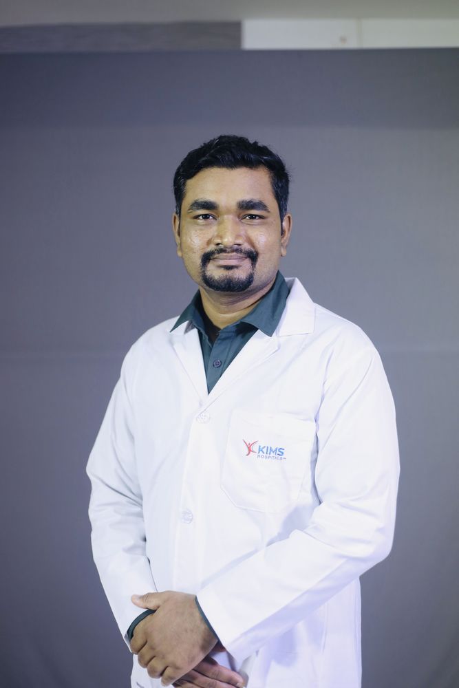
Dr. Manjunath M Negigoudara Transplant Pulmonologist · KIMS Hospital, Electronic City, Bengaluru
Home / Procedures / EBUS-TBNA in Bangalore: sampling lymph nodes without surgery
Procedure · Endobronchial ultrasoundAn ultrasound probe on the tip of a bronchoscope makes the airway wall transparent, so lymph nodes on the other side can be seen and a needle guided into them while you watch. It replaced a neck operation for most patients. Performed by Dr. Manjunath M Negigoudara at KIMS Hospital, Electronic City, Bengaluru.
Some of the most important structures in the chest sit just outside the airways. Lymph nodes in the mediastinum, the space between the lungs, collect drainage from the entire chest, which is why they enlarge in lung cancer, tuberculosis, sarcoidosis and lymphoma alike. A scan can show that a node is enlarged. It cannot reliably say why.
Until recently, getting tissue from those nodes meant mediastinoscopy: general anaesthesia, an incision at the base of the neck, and an instrument passed down behind the breastbone. Endobronchial ultrasound changed that. A small ultrasound probe built into the tip of a bronchoscope images through the airway wall, showing the nodes and the blood vessels around them, and a needle is passed through the wall into the node under continuous ultrasound vision.
Why this matters most in India
Enlarged mediastinal lymph nodes in an Indian patient can be tuberculosis, sarcoidosis, lymphoma or cancer, and treating the wrong one wastes months. EBUS samples are sent for cytology, for tuberculosis testing including molecular assays and culture, and for lymphoma workup, so a single procedure can separate diagnoses that look identical on a CT scan.
Whether cancer has reached the mediastinal lymph nodes, and which ones, determines whether surgery is worthwhile or whether chemotherapy, radiotherapy or immunotherapy should come first. Getting this wrong in either direction is serious: a patient can be denied a curative operation they could have had, or be put through major surgery that was never going to cure them. EBUS samples the node stations that matter and provides tissue for the molecular and immunotherapy marker testing that modern lung cancer treatment depends on.
A patient found to have mediastinal lymphadenopathy on a scan done for another reason needs an answer rather than a period of watchful waiting, particularly where tuberculosis is common. EBUS provides that answer in a day case.
Needle sampling of mediastinal nodes has become the preferred way to confirm sarcoidosis, outperforming the older approach of taking blind biopsies of lung tissue. Confirmation matters because sarcoidosis is treated quite differently from the conditions it mimics, and because many patients with it need no treatment at all. See interstitial lung disease.
A lesion adjacent to a central airway can be sampled through the wall under ultrasound guidance instead of by a needle passed through the chest wall, which avoids the risk of a collapsed lung that comes with the percutaneous route.
Preparation is the same as for a standard bronchoscopy: six hours without food, two without fluids, an individual plan for any blood thinners, your CT and PET images brought with you, and an escort to take you home afterwards. Full details are on the bronchoscopy page.
The scope is passed through the mouth under sedation with the airway numbed. The operator works systematically through the node stations, imaging each with ultrasound, measuring it, and checking the surrounding vessels with Doppler before any needle is passed. A needle is then advanced through the airway wall into the node while the ultrasound image shows it entering, and several passes are made per station.
Where rapid on-site evaluation is available, a smear from each pass is examined immediately so the operator knows whether the material is adequate before moving on. The sampling order follows staging principles, working from the stations that would indicate more advanced disease toward those that would indicate less, so that contamination cannot upstage a patient incorrectly.
A typical procedure takes 30 to 45 minutes. Recovery and discharge are as for a standard bronchoscopy, usually the same day.
Telling the laboratory what you are looking for before the procedure changes how samples are collected and preserved. This is one of the practical reasons the same clinician planning the case and performing it produces better results than splitting the two.
A positive result is reliable. If malignant cells, granulomas or tuberculosis are found in the node, that is your diagnosis and treatment can begin.
A negative result needs interpreting in context. If the nodes looked highly suspicious on PET or CT and the needle found nothing, the possibility remains that the involved part of the node was missed. In that situation surgical sampling may still be recommended. Your specialist should tell you before the procedure whether a negative result would end the investigation or lead to a further step, so that the plan is clear from the outset rather than being explained afterwards.
EBUS is an operator-dependent procedure. Knowing which stations to sample and in which order, recognising a node worth sampling from one that is not, avoiding vessels, and obtaining enough material for the tests that will actually be requested are all judgement rather than technique.
Dr. Manjunath M Negigoudara holds a Fellowship in Interventional Pulmonology from Manipal Hospitals, alongside MD in Pulmonary Medicine and DNB in Respiratory Medicine, and performs the full range of diagnostic and therapeutic airway procedures at KIMS Electronic City. See the interventional pulmonology service.
KIMS Hospital is on Hosur Road opposite the PES University EC Campus, about 15 minutes from Silk Board on the elevated expressway. Patients attend from HSR Layout, Koramangala, Sarjapur Road, BTM Layout, Jayanagar, JP Nagar and Bannerghatta Road, and from Bommasandra, Anekal, Chandapura and Attibele. You will need someone to take you home after sedation.
Patients travel for EBUS from Mysuru, Mangaluru, Hubballi-Dharwad, Belagavi, Kalaburagi, Shivamogga, Tumakuru, Davangere and Ballari, and from Chennai, Coimbatore, Madurai, Salem, Hyderabad, Warangal, Vijayawada, Visakhapatnam, Tirupati, Kochi, Thiruvananthapuram, Kozhikode, Goa, Mumbai, Pune, Nagpur, Ahmedabad, Delhi NCR, Kolkata, Bhubaneswar, Guwahati, Lucknow, Patna, Ranchi and Raipur. Because a staging decision often needs to be made quickly, send your CT and PET images on WhatsApp in advance so the consultation and the procedure can be scheduled on consecutive days. Kempegowda International Airport is about 45 km from the hospital.
Patients are seen from Bangladesh, Nepal, Sri Lanka, the Maldives and Bhutan, from Oman, the UAE, Saudi Arabia, Iraq, Yemen, Kuwait, Bahrain and Qatar, from Kenya, Tanzania, Ethiopia, Uganda, Rwanda, Sudan, Nigeria and Ghana, and from Myanmar, Indonesia and Malaysia. Imaging is reviewed over WhatsApp first, a written opinion can support a medical visa application, and diagnosis and staging are usually completed within a three to four day visit.