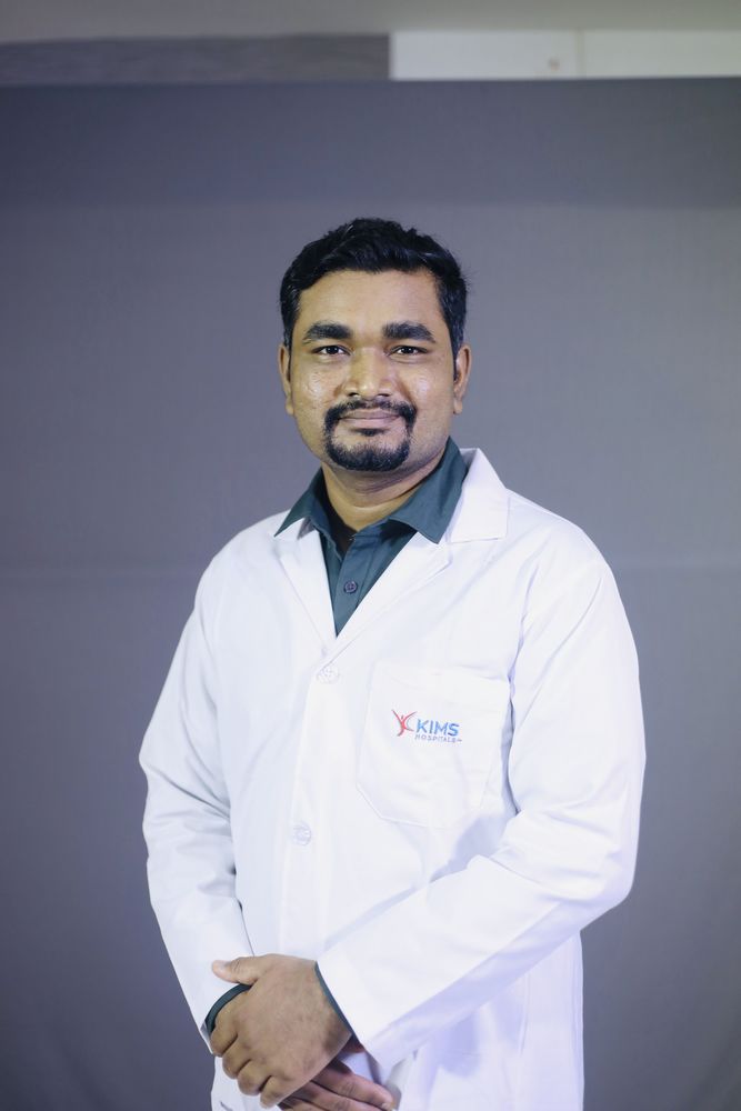
Dr. Manjunath M Negigoudara Transplant Pulmonologist · KIMS Hospital, Electronic City, Bengaluru
Home / Treatments / Interventional pulmonology at KIMS Electronic City
Service · Interventional pulmonologyBronchoscopy, airway stenting, EBUS-TBNA, cryotherapy, medical thoracoscopy and the full interventional pulmonology toolkit at KIMS Hospital, Electronic City - performed in-house by Dr. Manjunath M Negigoudara, FIP-trained (Fellowship in Interventional Pulmonology, Manipal Hospitals). Post-transplant airway management is a particular specialty.
Interventional pulmonology is the sub-specialty that uses bronchoscopy and minimally-invasive techniques to diagnose and treat diseases of the airways, lungs and pleura - conditions that historically required open thoracic surgery. It sits at the intersection of pulmonology, anaesthesia, thoracic surgery and radiology.
Dr. Manjunath M Negigoudara is FIP-trained (Fellowship in Interventional Pulmonology) from Manipal Hospitals, in addition to his transplant pulmonology practice at KIMS Hospital, Electronic City. This dual training matters most for post-lung-transplant patients - the same clinician who manages the transplant medically also performs the bronchoscopy, cryotherapy, dilatation, and stenting when complications arise. No handoff between teams, no delay for a visiting specialist.
The workhorse diagnostic procedure. A thin flexible scope is passed into the airways under sedation to inspect them, take biopsies, perform bronchoalveolar lavage and retrieve small foreign bodies. Usually a day case. Full detail on preparation, sedation, risks and recovery.
A wider metal scope used under general anaesthesia, for therapeutic work needing a controlled airway: central airway obstruction, major bleeding, large foreign bodies, tumour debulking and stent placement. Full detail on indications, stents and what the day involves.
Ultrasound on the scope tip lets a needle reach mediastinal and hilar lymph nodes through the airway wall under real-time guidance, replacing mediastinoscopy for most patients. Used to stage lung cancer and to diagnose sarcoidosis, tuberculosis and lymphoma. Full detail on the procedure and how results are interpreted.
A cryoprobe is passed through a flexible bronchoscope, cooled to freezing temperatures, and used to obtain lung tissue samples. Cryobiopsy gives larger, better-preserved specimens than standard transbronchial forceps biopsy - useful for diagnosing interstitial lung disease (IPF, NSIP, HP, connective tissue-related ILD) without the need for surgical VATS biopsy.
First-line treatment for symptomatic airway narrowing (post-tracheostomy stenosis, post-transplant anastomotic stenosis, benign or malignant tumour-related stenosis). A controlled-radial-force balloon is inflated across the narrowing for 60-90 seconds. Often needs 2-3 sessions but avoids the long-term issues of stents.
Freezes tissue rapidly to ablate lesions with minimal collateral damage. Preferred over electrocautery in transplanted airways because it preserves the cartilaginous framework and produces cleaner re-epithelialisation. Also used for mucus plug removal, blood clot extraction, and small tumour debulking.
For tumour ablation, granulation tissue removal, and bleeding control. Used cautiously in transplanted tissue where thermal injury has a higher risk of anastomotic breakdown.
| Stent type | Best for | Trade-off |
|---|---|---|
| Silicone (Dumon) | Benign stenosis expected to resolve; malacia in stable positioning | Removable + less granulation, but needs rigid bronchoscopy to place; mucus impaction more common |
| Self-expanding metallic (uncovered) | Malignant stenosis; refractory benign cases where permanent placement is acceptable | Placed via flexible bronchoscopy + low migration, but very difficult to remove; granulation grows through mesh |
| Covered / hybrid | Post-transplant dehiscence; recurrent stenosis needing barrier | Combines wall integration + granulation barrier, but higher migration risk |
Airway complications affect roughly 10-15% of lung transplant recipients per the ISHLT 2018 consensus. Five patterns dominate: anastomotic stenosis (most common late), dehiscence (first 4 weeks), bronchomalacia (dynamic collapse, 3-12 months), endobronchial infection (colonisation with Pseudomonas / S. aureus / Aspergillus / Candida), and granulation tissue.
Dr. Manjunath M Negigoudara’s FIP training + transplant pulmonology dual expertise means these complications are managed in-house at KIMS Electronic City without fragmenting the patient’s care between teams. See the detailed clinical guide: Post-lung-transplant airway interventions - a clinician’s guide.
Medical thoracoscopy (pleuroscopy) for pleural biopsy and management of recurrent pleural effusion, spontaneous pneumothorax, and pleural thickening. Performed under conscious sedation in the endoscopy suite - less invasive than surgical VATS but with diagnostic yield close to surgical biopsy for most pleural conditions.
Single-clinician continuity
Most lung transplant programmes refer post-transplant airway complications to a separate interventional pulmonology team. That fragments care. At KIMS Electronic City, the same clinician (Dr. Manjunath M Negigoudara) manages the transplant medically AND performs the bronchoscopy / cryotherapy / dilatation / stenting - no handoff, no delay, no loss of clinical context.
Referrals accepted for: post-transplant airway complications, difficult stent management, central airway obstruction, mediastinal lymphadenopathy for EBUS-TBNA, complex ILD needing cryobiopsy, recurrent pleural effusion.
Share imaging (HRCT chest, CT with contrast if oncological) + prior bronchoscopy reports via WhatsApp for pre-review before travelling to Bengaluru.
Call +91 79937 41199 · WhatsApp · Book online. Also see: transplant pulmonologist in Bangalore, best pulmonologist Electronic City.