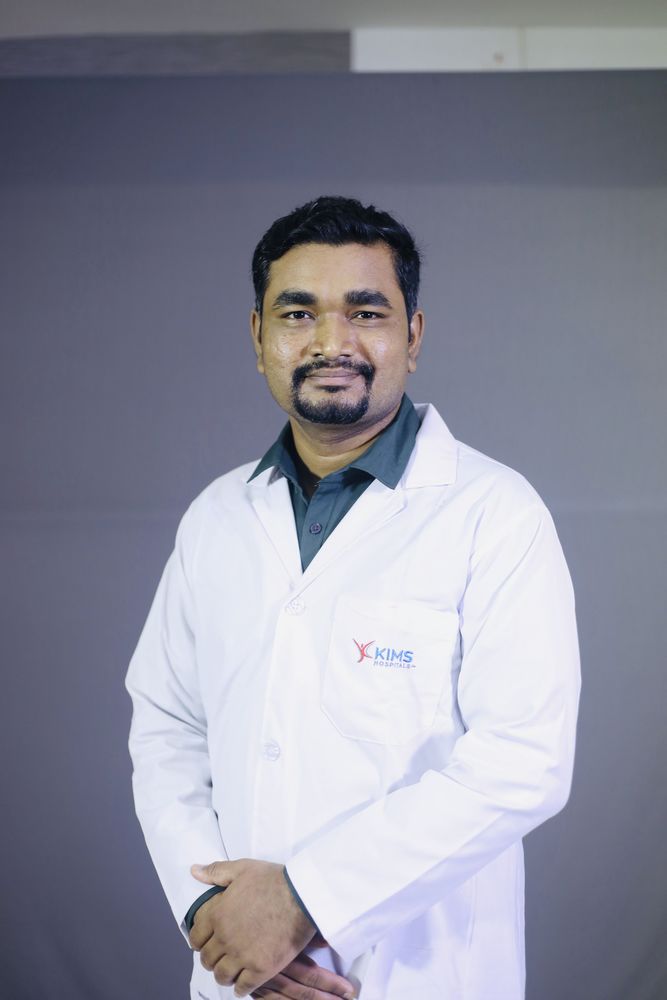
Dr. Manjunath M Negigoudara Transplant Pulmonologist · KIMS Hospital, Electronic City, Bengaluru
Home / Procedures / Bronchoscopy in Bangalore: what it is and what to expect
Procedure · Diagnostic bronchoscopyA thin flexible camera passed into the airways to see, wash and sample the lung directly. It is the workhorse diagnostic procedure in respiratory medicine, and for most patients it is a day case done under sedation. Performed by Dr. Manjunath M Negigoudara at KIMS Hospital, Electronic City, Bengaluru.
A bronchoscope is a flexible tube a little thicker than a pencil lead, carrying a camera and a working channel. Passed through the nose or mouth and down past the vocal cords, it allows the airways to be inspected directly and lets the operator wash, brush, biopsy or clear them. Almost everything respiratory medicine needs to know that a scan cannot tell it comes from this procedure.
The main reasons a pulmonologist recommends one are to find out what is causing shadowing on a scan, to obtain samples when infection is suspected and sputum has not given an answer, to investigate coughing blood, to sample abnormal tissue or lymph nodes, and to clear an airway that is blocked by secretions, a clot or a foreign body.
Three different procedures, three different pages
This page covers flexible diagnostic bronchoscopy. For sampling lymph nodes with ultrasound guidance, see EBUS-TBNA. For a blocked or collapsing central airway, bleeding, a large foreign body or stent placement, see rigid bronchoscopy.
Sterile saline is instilled into a chosen segment and suctioned back, returning cells and organisms from the small airways and air sacs. The fluid goes for differential cell counts, microbiology including tuberculosis and fungal studies, and cytology. In interstitial lung disease the cell pattern helps separate hypersensitivity pneumonitis and sarcoidosis from other causes. In immunosuppressed patients, including lung transplant recipients, it is often the only way to identify the organism responsible for a new infiltrate.
Visible abnormal tissue in an airway can be biopsied directly. Tissue deeper in the lung is sampled by passing forceps through the airway wall into the surrounding lung, usually with fluoroscopic guidance. Transbronchial biopsy is particularly useful in sarcoidosis, in suspected malignancy, and in transplant recipients where it remains the standard way of assessing rejection.
A probe is cooled so tissue freezes to its tip and is removed as a single larger fragment. The samples are bigger and less crushed than forceps biopsies, which matters to a pathologist trying to classify interstitial disease. The trade-off is a higher bleeding risk, so it is performed with a protected airway and bleeding control immediately available. For many patients it now replaces surgical lung biopsy, which requires general anaesthesia and a chest drain.
Thick secretions and mucus plugs causing lobar collapse can be suctioned and washed out, often with immediate improvement in oxygenation. Small foreign bodies can be retrieved, though larger ones and anything in a child are safer handled with a rigid scope.
A cannula is placed and monitoring attached for oxygen saturation, heart rhythm and blood pressure. Local anaesthetic is sprayed onto the throat and sedation given through the drip. Oxygen is delivered throughout. The scope passes through the nose or mouth, past the vocal cords and into the airways, and the planned samples are taken. Most patients are drowsy and recall little.
A straightforward diagnostic procedure takes 20 to 30 minutes. You are then observed until the sedation wears off, which is typically two to four hours. After a transbronchial biopsy or cryobiopsy a chest X-ray is usually taken to check for a collapsed lung before discharge. You must not eat or drink until the throat anaesthetic has worn off, generally about two hours, because swallowing is not safe until sensation returns.
Expect a sore throat, a hoarse voice and a cough for a day or so. Small streaks of blood in the sputum for a day after biopsy are expected. A mild fever on the evening of a lavage is a known reaction and settles.
Seek urgent attention for breathlessness that is new or worsening, chest pain that is sharp and worse on breathing in, coughing more than a tablespoon of fresh blood, or fever above 38.5 degrees persisting beyond 24 hours. Call +91 79937 41199 or attend the KIMS Electronic City emergency department.
For inspection and washings alone, complications are uncommon. Where biopsies are taken, two matter. Bleeding is usually minor and controlled during the procedure, but is greater with cryobiopsy and in patients with pulmonary hypertension or clotting problems. A collapsed lung, pneumothorax, occurs in a minority after transbronchial biopsy and sometimes needs a chest drain, which is why a post-procedure X-ray is routine.
Less common are transient falls in oxygen levels, heart rhythm disturbance, and reaction to sedation. Serious complications are rare. Each is discussed in the consent conversation before the procedure, with the specific risk for your circumstances rather than a generic figure.
The decision that most affects a bronchoscopy result is made before the scope is picked up: which technique will answer this question in this patient, and which segment to sample. A lavage from the wrong lobe, or forceps where a cryobiopsy was needed, produces a non-diagnostic result and the patient goes through the procedure again.
Dr. Manjunath M Negigoudara holds a Fellowship in Interventional Pulmonology from Manipal Hospitals alongside MD in Pulmonary Medicine and DNB in Respiratory Medicine. Because he also runs the transplant service, diagnostic bronchoscopy, therapeutic airway work and the transplant pathway sit in one department rather than three. Read more about the interventional pulmonology service.
KIMS Hospital is on Hosur Road opposite the PES University EC Campus, about 15 minutes from Silk Board on the elevated expressway. Day-case bronchoscopy patients attend from HSR Layout, Koramangala, Sarjapur Road, BTM Layout, Jayanagar, JP Nagar and Bannerghatta Road, and from Bommasandra, Anekal, Chandapura and Attibele. Remember that you need someone to take you home.
Patients travel for bronchoscopy from Mysuru, Mangaluru, Hubballi-Dharwad, Belagavi, Kalaburagi, Shivamogga, Tumakuru, Davangere and Ballari, and from Chennai, Coimbatore, Madurai, Salem, Hyderabad, Warangal, Vijayawada, Visakhapatnam, Tirupati, Kochi, Thiruvananthapuram, Kozhikode, Goa, Mumbai, Pune, Nagpur, Ahmedabad, Delhi NCR, Kolkata, Bhubaneswar, Guwahati, Lucknow, Patna, Ranchi and Raipur. Send your CT images on WhatsApp beforehand so the procedure can be planned and, where appropriate, the consultation and bronchoscopy scheduled on consecutive days rather than requiring two trips. Kempegowda International Airport is about 45 km away.
Patients are seen from Bangladesh, Nepal, Sri Lanka, the Maldives and Bhutan, from Oman, the UAE, Saudi Arabia, Iraq, Yemen, Kuwait, Bahrain and Qatar, from Kenya, Tanzania, Ethiopia, Uganda, Rwanda, Sudan, Nigeria and Ghana, and from Myanmar, Indonesia and Malaysia. Scans and reports are reviewed over WhatsApp first, a written opinion can support a medical visa application, and diagnostic work is usually compressed into a three to four day visit with video follow-up afterwards.